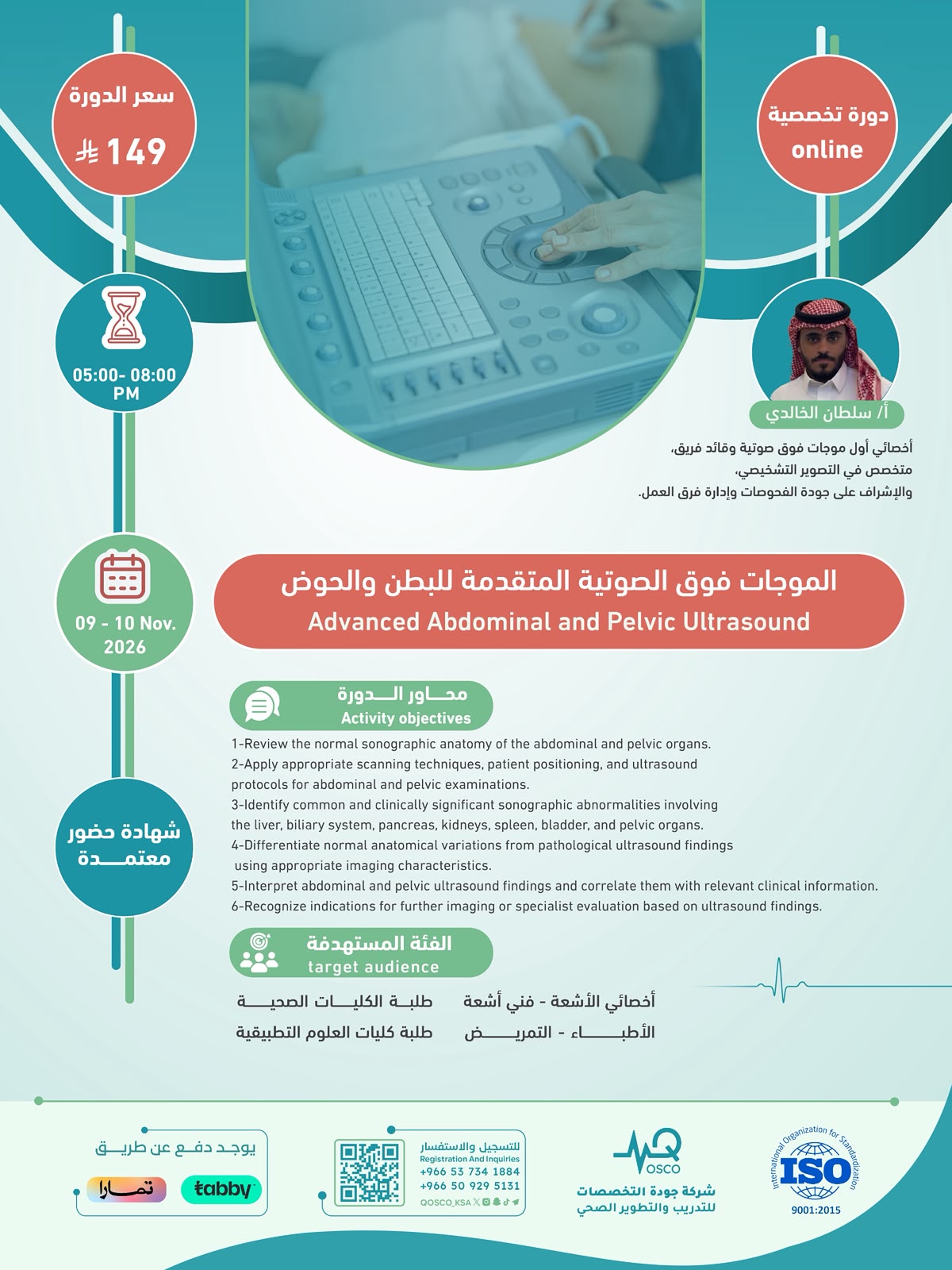
الموجات فوق الصوتية المتقدمة للبطن والحوض -1️⃣1️⃣
تفاصيل الدورة
-
ما سوف تتعلم فى هذه الدورة
1-Review the normal sonographic anatomy of the abdominal and pelvic organs.
2-Apply appropriate scanning techniques, patient positioning, and ultrasound protocols for abdominal and pelvic examinations.
3-Identify common and clinically significant sonographic abnormalities involving the liver, biliary system, pancreas, kidneys, spleen, bladder, and pelvic organs.
4-Differentiate normal anatomical variations from pathological ultrasound findings using appropriate imaging characteristics.
5-Interpret abdominal and pelvic ultrasound findings and correlate them with relevant clinical information.
6-Recognize indications for further imaging or specialist evaluation based on ultrasound findings.
عن الدورة
- التصنيف دورات أونلاين ( تخصصية ) معتمدة من شركة جودة التخصصات بشهادة حضور
- السعر 149 ريال سعودى
-
- اللغة العربية و الانجليزية
- المدة2 ايام
- شهادة اتمام الدورة