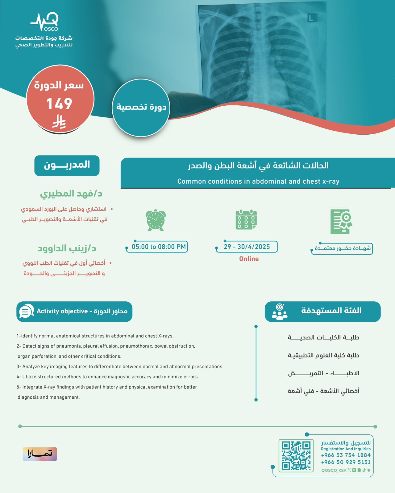
الحالات الشائعة في اشعة البطن والصدر - 4️⃣

تفاصيل الدورة
-
ما سوف تتعلم فى هذه الدورة
1-Identify normal anatomical structures in abdominal and chest X-rays.
2- Detect signs of pneumonia, pleural effusion, pneumothorax, bowel obstruction, organ perforation, and other critical conditions.
3- Analyze key imaging features to differentiate between normal and abnormal presentations.
4- Utilize structured methods to enhance diagnostic accuracy and minimize errors.
5- Integrate X-ray findings with patient history and physical examination for better diagnosis and management.
عن الدورة
- التصنيف دورات أونلاين ( تخصصية ) معتمدة من شركة جودة التخصصات بشهادة حضور
- السعر 149 ريال سعودى
- اللغة العربية - الانجليزية
- المدة2 ايام
- شهادة اتمام الدورة